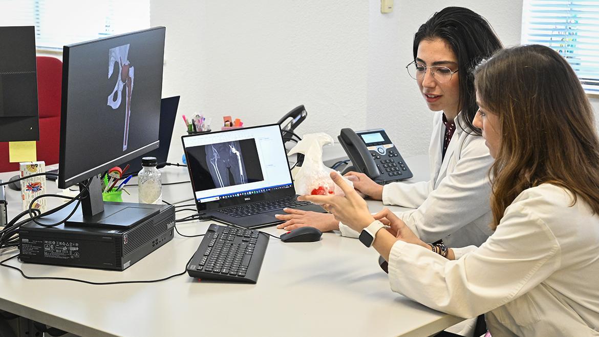
Surgical planning
We use digital tools and three-dimensional reconstructions to study each patient's anatomy and support the preparation of complex surgical procedures.
DISCOVER MORE →
At UPAM3D, we combine medicine, engineering and 3D technology to bring personalised planning and manufacturing into the hospital.
We study anatomy in 3D to support the preparation of surgical procedures.
We create anatomical models and solutions tailored to each clinical case.
Science and technology working together to improve healthcare.
Discover how technology and engineering help us better understand clinical cases and develop solutions tailored to patients' needs.
We use digital tools and three-dimensional reconstructions to study each patient's anatomy and support the preparation of complex surgical procedures.
DISCOVER MORE →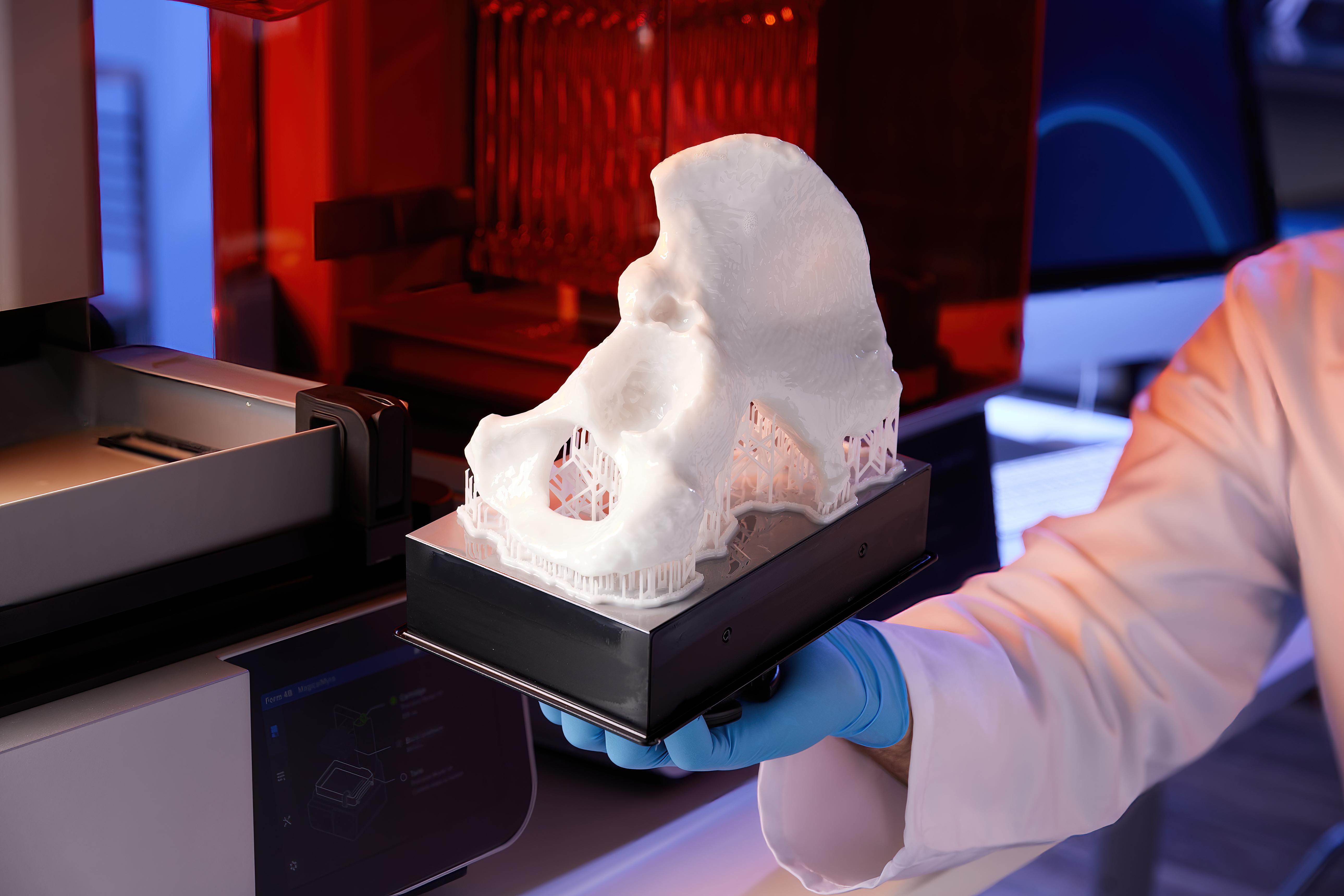
We design and manufacture anatomical models, surgical guides and personalised solutions for a wide range of clinical needs.
DISCOVER MORE →
We take part in research projects and training activities to explore new applications of advanced planning and manufacturing technologies.
DISCOVER MORE →Our unit brings together different areas of expertise, working in coordination to turn a clinical need into a personalised solution.
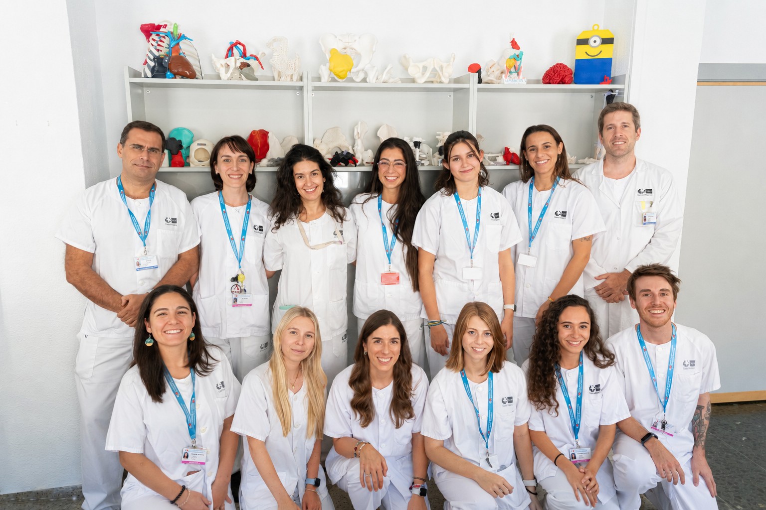
Technical design and development
Clinical and biomedical engineering supports the development of personalised solutions for different healthcare needs.
Behind every project are professionals from different disciplines who share knowledge, ideas and experience to address challenges in healthcare.
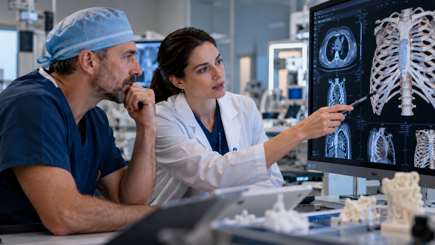
MEET OUR TEAM →Every project begins with a clinical need. Professionals from different disciplines work together to study the case, develop a solution and evaluate the result.
Everything begins with a need identified by healthcare professionals. We study the case, discuss the requirements and explore how technology might help.
Select a technology and try a short interactive demonstration. These are simplified educational activities, not real clinical simulations.
Augmented reality can overlay digital anatomical information on a view of the patient.
Drag the digital liver onto the marked area of the abdomen.
Simplified educational representation.
Explore how medical images can be transformed into a digital anatomical model and, eventually, a physical 3D print.
A CT scan produces a series of cross-sectional images of the body. Move the slider to explore different slices.
Medical images can be segmented to create a three-dimensional representation of the anatomy.
The digital anatomical model is ready for the next step.
Start a simplified simulation of layer-by-layer manufacturing.
You have explored the journey from medical images to a physical anatomical model.
Educational simulation only. No real patient data are used.
Electronic sensors and devices are essential in modern healthcare. A pulse oximeter estimates blood oxygen saturation and measures pulse rate.
Drag the finger into the highlighted opening. The device will display an illustrative measurement.
Real pulse oximeters use optical sensors to estimate blood oxygen saturation.
Fictional values. Not a medical measurement.
Study a simplified CT scan, identify a lesion and explore possible routes through the image.
Look at the scan and click on the marked area in the liver.
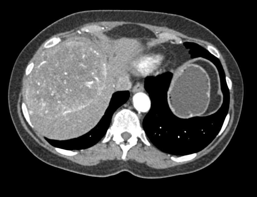
Choose the route that avoids crossing the highlighted blue structure in this simplified illustration.
You have identified the lesion and selected a route in this educational exercise.
Educational simulation only. Not suitable for clinical decisions.
Adjust the dimensions of a virtual prosthesis to match the target outline.
Use the controls to match the target dimensions.
Adjust the width and length until the shape matches the red target outline.
This simulation uses fictional dimensions and is not a medical device design tool.
Hydrogels can retain large amounts of water within their structure. Explore how changes in experimental conditions may affect a virtual sample.
Choose an alginate concentration and a calcium chloride exposure time.
Change the concentration and exposure time to explore a virtual gelation experiment.
This is an educational representation, not a predictive model of real hydrogel behaviour. Actual gelation depends on formulation and experimental conditions.
Three ways 3D technology is integrated into hospital practice.
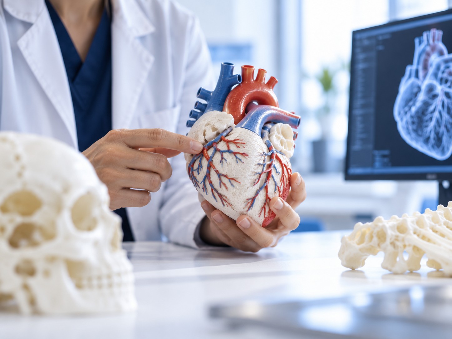
Visualise complex structures in a clear, tangible way.
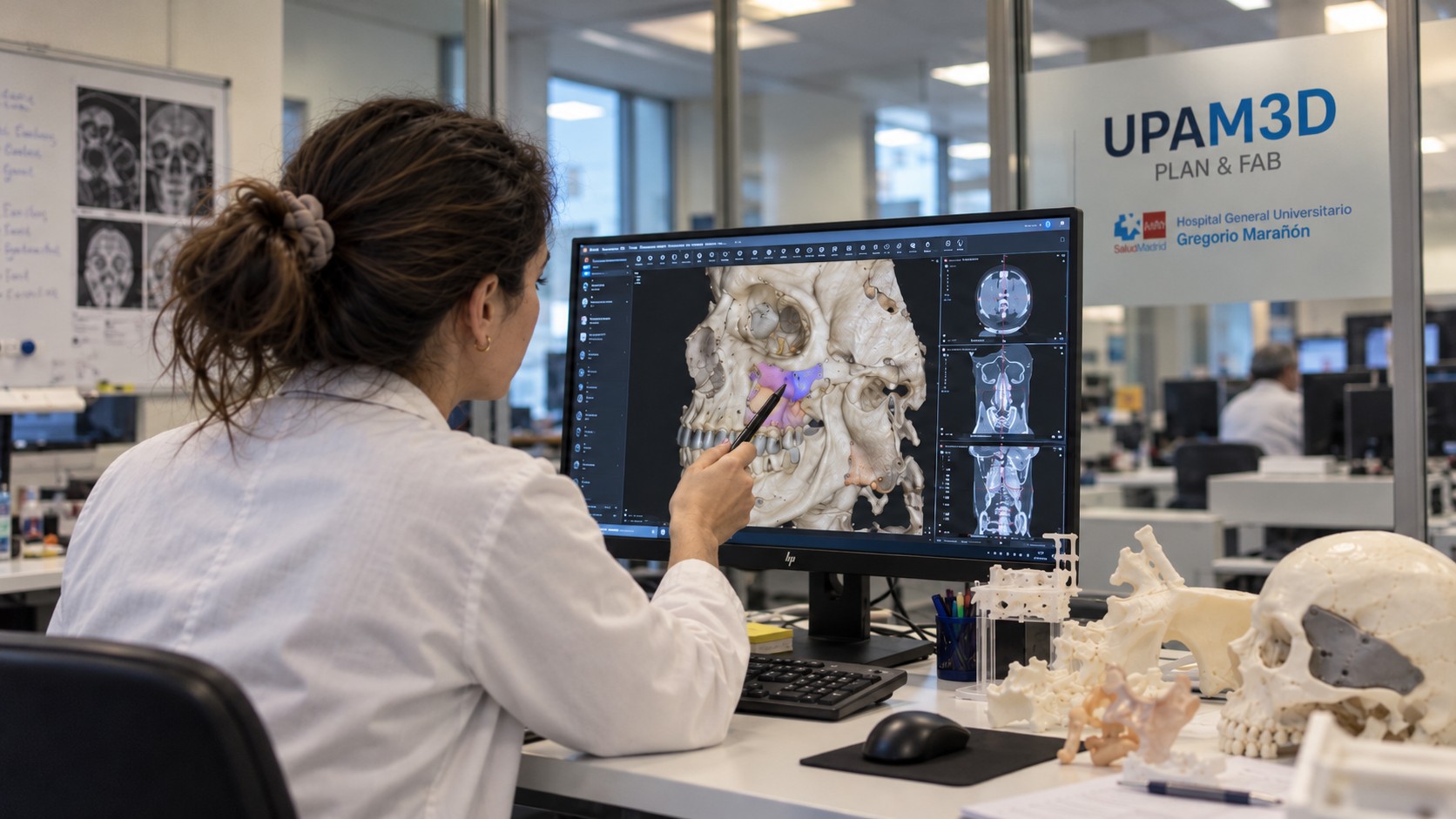
Support the study of clinical cases using digital tools.

Explore new ways to teach and understand anatomy.
Discover how we use 3D technology to support surgical planning and develop solutions tailored to individual patients.
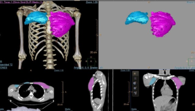
3D technology helped the team study anatomical differences and prepare a reconstruction tailored to the patient's chest.
EXPLORE THE CASE →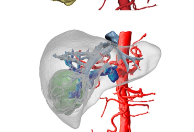
A 3D digital model helped the medical team study the liver and its blood vessels before a complex procedure.
EXPLORE THE CASE →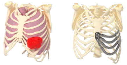
Advanced planning and 3D printing supported the design of a solution adapted to the patient's anatomy.
EXPLORE THE CASE →Learn how the unit is organised and the quality framework under which it operates. Meet the professionals behind UPAM3D on Our team .
Associated resources: 3D Bone Bank, 3D Hybrid Operating Room and point-of-care manufacturing.
Quality management system certified to ISO 13485:2018, in accordance with European Regulation (EU) 2017/745 and Spanish Royal Decree 192/2023.
Custom-made medical device manufacturer licence granted in 2020 and extended in 2023.
Download AENOR certificate (Spanish PDF) →Discover what we do and how we use technology in healthcare, explained in simple terms.
3D printing makes it possible to manufacture anatomical models and certain personalised solutions. These models can help healthcare professionals understand complex structures, prepare for surgical procedures, support training and improve clinical communication.
An anatomical model is a physical or digital representation of an anatomical structure. It can be created from medical images and used to help study anatomy or plan certain medical procedures.
Augmented reality combines digital images or models with a view of the real world. It can help visualise anatomical structures and explore new approaches to planning, training and visual support.
An anatomical model represents a structure of the human body and can be used for visualisation or study. A personalised prosthesis, on the other hand, is a device designed to perform a specific function and meet the needs of an individual patient. Where applicable, its development is subject to specific safety and medical device regulatory requirements.
Whether you have a question, a collaboration proposal or would simply like to know more about our work, we would love to hear from you.
Contact UPAM3D through any of the following channels.